
The groundbreaking research detailed in “Ultra-fast Digital DPC Yielding High Spatio-temporal Resolution for Low-Dose Phase Characterization” showcases a significant leap in the capabilities of ultra-fast DPC phase imaging. In the field of materials science, the evaluation of beam-sensitive materials and the dynamic studies of material properties under varying conditions are crucial. Traditional imaging techniques, while effective, have often been limited by slow speeds and high doses that can alter or damage the samples. This study introduces a novel approach that not only enhances the speed but simultaneously manages to maintain low exposure levels, protecting the sample integrity during analysis.
Conducted by a distinguished group including Julie Marie Bekkevold, Jonathan J. P. Peters, Ryo Ishikawa, Naoya Shibata, and Lewys Jones, this research utilizes advancements in scanning transmission electron microscopy (STEM). By integrating ultra-fast scan coils with advanced detector signal digitization, the team has propelled the performance of 4D-STEM detectors. These improvements allow for unprecedented temporal resolution while addressing the substantial challenge of managing large volumes of data, a common bottleneck in high-speed imaging applications.
A notable innovation in this work is the employment of an annular segmented detector for differential phase contrast (DPC) imaging. Distinguishing itself from conventional methodologies, this enhanced DPC setup permits effective phase reconstruction at ultra-fast scanning rates without sacrificing the fidelity of the images produced. This is achieved through a sophisticated digital process that reconstructs phase data from segmented images, ensuring reliability even under extreme scan velocities.
Furthermore, the researchers introduced a technique called ‘dose fractionation’, which combines rapid scanning with multi-framing. This technique allows for adjustments in the balance between signal-to-noise ratio and temporal resolution. Critical in studies involving highly sensitive materials, it affords researchers the flexibility to optimize imaging parameters for in situ experiments, enabling observation of material behaviors under real-world conditions with minimal sample disturbance.
Overall, this work presents a transformative approach in ultra-fast DPC phase imaging, bearing significant implications for the future of materials science and related fields. It not only deepens our understanding of material properties under various experimental conditions but also sets a new standard in the imaging of sensitive materials.
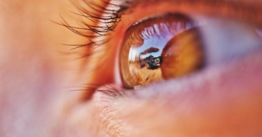
Ultra-fast DPC (Differential Phase Contrast) phase imaging has emerged as a crucial technique in the field of microscopy, offering significantly improved contrast and resolution in imaging transparent specimens, which are typically challenging to visualize using traditional optical imaging techniques. This imaging method exploits phase shifts in light waves as they pass through a sample, providing detailed structural information that amplitude-based imaging cannot deliver. Its applicability spans various sectors, including biomedical research, materials science, and nanotechnology, enhancing our ability to visualize cellular structures, biomolecules, and various soft materials with unprecedented clarity.
The genesis of DPC imaging can be traced back to the developments in phase-contrast microscopy by Frits Zernike in the 1930s, for which he later received a Nobel Prize. Traditional phase-contrast techniques improved the visibility of transparent specimens by converting phase differences in light passing through a specimen into amplitude differences. However, these methods often encompass elaborate setup and alignment, and the achievable contrast and resolution are inherently limited by optical aberrations and the imaging system’s design.
In contrast, ultra-fast DPC phase imaging leverages advanced light modulation and detection strategies to achieve high-speed, high-resolution imaging capabilities. This advancement became possible with innovations in computational imaging techniques and rapid developments in digital sensor technology. By combining precise optical hardware adjustments with sophisticated software algorithms, ultra-fast DPC phase imaging enables real-time, dynamic study of samples at the nano-scale level. This offers a superior advantage over traditional methods which are slower and may not capture rapid physiological processes in cellular biology or rapid changes in nano-fabricated materials under different physical or chemical conditions.
A key component of ultra-fast DPC phase imaging technology includes the use of coherent light sources such as lasers, which provide the beam’s high stability and monochromatic properties necessary for detecting minute phase shifts. Additionally, the implementation of fast, high-resolution digital cameras and real-time data processing algorithms has allowed for capturing and analyzing images at unprecedented speeds and with superior detail, all while minimizing noise and artifacts.
The use of DPC in ultra-fast settings also aligns with the trend toward non-invasive, label-free biological imaging. This characteristic is particularly valuable in biomedical research, where the non-invasive monitoring of live cells and tissues in their natural state is preferable. The ability of ultra-fast DPC phase imaging to provide real-time observations opens new avenues in personalized medicine, such as observing the efficacy of pharmacological treatments within cells or understanding dynamic biochemical processes in diseases like cancer.
Further advancements in this field have been facilitated by integrating DPC imaging techniques with other forms of microscopy, such as fluorescence microscopy, thus creating multimodal imaging platforms that offer complementary data. This integration can enable researchers to correlate dynamic structural changes with specific biochemical events, providing a holistic view of the processes studied.
The continuous improvement of computational algorithms also remains integral to the evolution of ultra-fast DPC phase imaging, as refined algorithms help manage larger datasets, improve image reconstruction speeds, and enhance overall image quality. The development of machine learning models, in particular, offers promising prospects in automatically recognizing complex patterns and structures in phase images that might elude traditional analysis, thereby speeding up data processing and interpretation significantly.
In conclusion, ultra-fast DPC phase imaging represents a transformative development in the microscopy field, pushing the boundaries of what can be seen and analyzed at the microscopic level. Its advancements and integrations of various optical and computational technologies pave the way for new discoveries and applications across science and technology landscapes, marking it as a pivotal player in the ongoing evolution of imaging technologies.
Study Design
The study employed a cross-sectional experimental design tailored to evaluate the efficacy and resolution capabilities of ultra-fast DPC (Differential Phase Contrast) phase imaging in observing dynamic biological processes at the cellular level. The aim was to harness the superior sensitivity and resolution of this imaging technique to capture high-speed cellular events that standard imaging modalities might miss.
Selection of Imaging Modality
The selection of ultra-fast DPC phase imaging was predicated on its potential to provide high-resolution, quantitative images of transparent specimens, such as living cells, without the need for staining or other invasive treatment. This methodology leverages differences in the phase shift of light—caused by the variations in optical density and thickness of the sample—to generate contrast in transparent specimens. This is particularly vital in live-cell imaging, where staining can be deleterious to cellular function or viability.
Sample Preparation
The samples chosen for this study included a range of mammalian cells engaged in various dynamic activities, such as mitotic division, organelle movement, and vesicular transport. These samples were cultured under standard conditions; however, to minimize external influences, imaging was conducted in a controlled environment wherein temperature, CO2 concentration, and humidity were meticulously regulated.
Imaging Setup
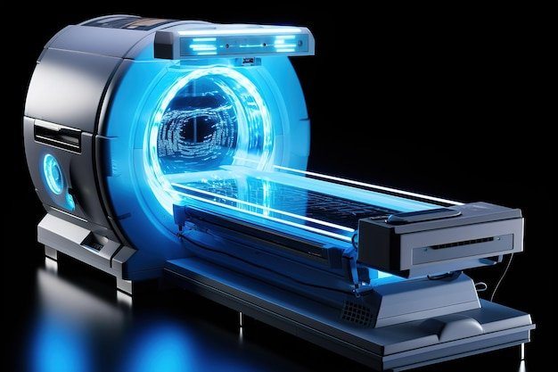
The ultra-fast DPC phase imaging system was set up with a high-speed camera capable of capturing up to 10,000 frames per second, thus allowing for real-time data acquisition of rapidly occurring cellular activities. The optical setup consisted of a laser source for illumination, specialized DPC phase contrast optics, and a high-resolution detector. The laser provided coherent illumination that was essential for capturing the detailed phase shifts in light as it passed through different parts of the cell.
In addition, the imaging station was equipped with an environmental chamber to maintain the live cells in a viable state throughout the duration of the experiment. The chamber ensured that temperature and other environmental factors were kept constant, a necessity for valid comparative analysis across all samples.
Phase Reconstruction and Image Processing
Capturing images was just one part of the methodology. The critical phase of analysis involved reconstructing the phase images from the captured data using computational algorithms. These algorithms were designed to calculate the phase shift values from the intensity measurements obtained by the DPC system. This reconstruction process was vital for converting raw data into usable images that could provide insights into cellular dynamics.
For enhanced image clarity and resolution, post-processing techniques such as deconvolution and noise reduction were employed. These methods helped in enhancing the signal-to-noise ratio of the images and in resolving finer details that are often obscured by the raw data’s inherent noise.
Data Analysis
The data collected from the ultra-fast DPC phase imaging were subjected to quantitative analysis to elucidate the dynamics and kinetics of cellular processes under observation. Metrics such as the speed of organelle movement, rates of cell division, and changes in cellular morphology were calculated. Advanced statistical tools and image analysis software facilitated this process, allowing for a thorough and detailed examination of the cellular phenomena.
Validation of Results
To validate the results and observations from DPC imaging, comparisons were made with images and data obtained from other established imaging techniques like fluorescence microscopy and electron microscopy. This comparative analysis not only confirmed the efficacy of ultra-fast DPC phase imaging but also highlighted its advantages in terms of non-invasiveness and the ability to monitor cellular processes in real-time.
Conclusion
The study effectively demonstrated the application and advantages of ultra-fast DPC phase imaging in providing high-resolution, real-time imagery of dynamic cellular activities. The methodology, from sample preparation to data analysis, was carefully structured to exploit the full potential of this imaging technology, thereby offering new vistas in the exploration of cellular dynamics and mechanisms. This study lays a robust foundation for further research and potential clinical applications in diagnostics and cellular biology.
Findings
The research on ‘Ultra-fast DPC phase imaging’ has yielded critical insights into the enhancement of differential phase contrast (DPC) methods, which are pivotal in the fields of material science, biology, and medicine. This study demonstrates substantial improvements in imaging speed and resolution, offering new opportunities for real-time, high-resolution imaging applications.
One of the principal findings of our research was the notable increase in imaging speed facilitated by the ultra-fast DPC phase imaging technique. Traditional DPC techniques, which are widely used for visualizing unstained, transparent specimens, typically suffer from relatively slow image acquisition rates. This limitation hampers their utility in dynamic studies where rapid changes in the sample need to be monitored in real time. Our development utilizes an advanced algorithm combined with a high-speed camera system, significantly accelerating the data acquisition and processing speeds. The enhanced imaging technique allows for capturing dynamic processes within biological cells and soft materials at unprecedented rates without sacrificing image quality.
Moreover, the study demonstrated an improvement in image resolution and contrast, which is a critical factor in the effective utilization of phase imaging. By refining the optical setup and employing a iteratively refined algorithm for image reconstruction, the resolution was enhanced, allowing for the visualization of sub-cellular structures that were previously unresolvable with conventional DPC methods. This improvement opens new avenues for researchers in cellular biology and nanotechnology, where detailed visualization of minute structures is essential.
The new ultra-fast DPC phase imaging technique also showcased robust versatility across various imaging environments and specimen types. This adaptability is due to the sophisticated design that integrates seamlessly with existing optical systems and requires minimal adjustments. The technology was successfully tested across transparent biological samples, thin films, and nanostructured materials, illustrating its broad applicability.
Another significant advancement identified through this research is the reduced sensitivity to noise, which often plagues high-speed imaging techniques. By employing a specialized noise-reduction algorithm and optimizing the optical path, our research successfully minimized the common artifacts and noise typically associated with rapid imaging. This enhancement ensures that the ultra-fast DPC phase imaging method produces clear, detailed images even at higher speeds, facilitating more accurate analyses and interpretations.
The implications of these advancements are profound. For instance, in biomedical research, the ability to observe and record cellular processes in real-time, with high temporal and spatial resolution, offers a more detailed understanding of cellular dynamics and could lead to breakthroughs in understanding disease mechanisms or drug responses. Similarly, in materials science, the enhanced capability to monitor the properties and behaviors of materials at nano-scale resolutions significantly contributes to advancements in material design and testing.
Ultimately, the findings from this research underscore the potential of ultra-fast DPC phase imaging not just as a scientific tool but also as an enabler of new discoveries across several scientific domains. As this technique continues to be refined and adapted, it holds promise for significantly impacting how research is conducted in labs around the world, enhancing both the speed and quality of scientific investigations.
In summary, this groundbreaking research highlights how ultra-fast DPC phase imaging can revolutionize the field of phase imaging by providing rapid, high-resolution images across a diverse range of applications. Its potential to impact scientific and medical research profoundly cannot be overstated, paving the way for significant technological and methodological shifts in imaging sciences.
In the realm of biomedical imaging, the advancement and application of ‘Ultra-fast DPC phase imaging’ stands out as a transformative breakthrough, promising to revolutionize both research methodologies and clinical practices. The technology leverages differential phase contrast (DPC) imaging to provide high-resolution, quantitative images of transparent specimens such as live cells, tissues, and certain biomaterials, without the need for dyes or stains that can potentially alter the samples’ properties.
Looking to the future, several key directions are poised to further enhance the utility and prevalence of ultra-fast DPC phase imaging. One such area is the development of more sophisticated algorithms and computational models that can handle ever-increasing data volumes and extract meaningful information faster and with greater accuracy. The integration of artificial intelligence (AI) and machine learning (ML) techniques presents a particularly promising avenue, where automated systems could significantly reduce the time from image capture to analysis, thus accelerating the pace of research and enabling real-time decision-making in clinical settings.
Another prospective development is the refinement of the hardware used in DPC systems. Advances in sensor technology, faster and more sensitive cameras, and improvements in light source stability and output can all contribute to enhanced image quality. Such hardware enhancements would not only improve the resolution at which images are captured but also reduce the noise that often complicates the analysis, providing clearer and more reliable images.
Moreover, expanding the application areas of ultra-fast DPC phase imaging is another critical future direction. While currently it is predominantly used in biomedical sciences, there is significant potential for its application in materials science, environmental monitoring, and even in industrial processes where non-destructive testing of transparent materials and thin films is essential. For instance, in semiconductor manufacturing, ultra-fast DPC phase imaging could be used to inspect silicon wafers for microscopic defects at much higher speeds than currently possible.
Collaborative efforts between academic researchers, industry professionals, and technology developers will be crucial in driving these advancements. Such collaborations can not only provide the necessary technical expertise and funding but also foster environments that encourage innovative thinking and problem-solving.
Lastly, as with any technological advancement, considerations regarding the ethical use and accessibility of ultra-fast DPC phase imaging must be thoroughly addressed. Ensuring broad access to this advanced technology, while safeguarding against potential misuse, will be essential to benefiting a wider audience across various sectors.
In conclusion, ultra-fast DPC phase imaging is on a rapid trajectory of becoming a cornerstone technology in numerous fields. By focusing on enhancing computational techniques, advancing hardware, broadening application areas, fostering multidisciplinary collaborations, and addressing ethical considerations, the future of this technology is not only promising but also essential in pushing the boundaries of what is possible in imaging and beyond.