
In the realm of orthodontics, understanding the mechanisms driving alveolar bone loss is critical. Recent research on GPX4 ferroptosis in orthodontic treatment reveals significant insights into the biological processes underpinning bone degradation. Specifically, this study by Wang Mengjia and colleagues explores the GPX4-mediated pathway of bone ferroptosis under mechanical stress and its impact on bone formation through the YAP-TEAD signalling pathway. The research posits that ferroptosis, a specialized form of cell death characterized by iron-dependent lipid peroxidation, plays a crucial role in the response of alveolar bone to mechanical loading during orthodontic tooth movement (OTM).
The study employs both in vitro and in vivo models to investigate the effects of mechanical force on osteoblast cellular behavior and ferroptosis induction. Using mouse osteoblasts and GPX4 knockdown cells, the research delineates how different intensities of compressive force stimulate distinct responses in terms of lipid peroxidation, ferroptosis marker expression, and osteogenesis. Moreover, the study leverages pharmacological intervention with ferrostatin-1, a known inhibitor of ferroptosis, to ascertain its protective role against bone loss.
The findings suggest a pivotal function of the GPX4 enzyme in modulating the ferroptosis pathway and link these cellular mechanisms to the broader YAP-TEAD signalling network, thereby providing a potential therapeutic target for mitigating bone loss in orthodontic settings. This study not only enhances our understanding of cell death in orthodontic treatment but also opens new avenues for clinical strategies to prevent bone degradation in patients undergoing orthodontic procedures.
Understanding the cellular responses to mechanical stress in orthodontic treatment is essential for improving outcomes and mitigating complications such as unwanted bone loss. Prior research into the effects of mechanical loading on alveolar bone has primarily focused on bone resorption and formation. However, recent advancements in cellular biology have illuminated the role of specialized cell death processes, such as ferroptosis, in orthodontic scenarios. The study on GPX4 ferroptosis in orthodontic treatment by Wang Mengjia and colleagues represents a fundamental shift towards examining the molecular pathways that underlie bone response to orthodontic forces.
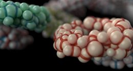
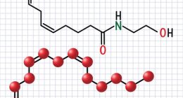
Ferroptosis is a type of programmed cell death distinctly characterized by iron-dependent lipid peroxidation, leading to severe cellular damage and death. The enzyme glutathione peroxidase 4 (GPX4) is crucial in inhibiting ferroptosis by converting lipid hydroperoxides into non-toxic lipid alcohols, thus preventing membrane damage. In the context of orthodontics, the activation of ferroptosis in alveolar bone cells can potentially lead to enhanced bone degradation, disrupting the balance required for optimal bone remodelling during tooth movement.
The probing into the GPX4-mediated ferroptosis during orthodontic tooth movement (OTM) marks a pioneering approach to understanding the biological adversity that can accompany mechanical stress in orthodontic care. By focusing on the GPX4 enzyme and its regulatory role in ferroptosis under these specific conditions, Wang Mengjia’s research critically links this cell death pathway with orthodontic-induced alveolar bone changes.
The involvement of the YAP-TEAD signalling pathway, known for its role in cell proliferation, apoptosis, and organ size regulation, adds another layer of complexity and relevance. The study reveals how mechanical force may trigger ferroptotic pathways in osteoblasts, negatively affecting their survival and function, thus impairing bone formation. Inhibiting ferroptosis through pharmacological means, such as with ferrostatin-1, showcases a promising avenue to safeguard osteoblasts and maintain bone integrity during orthodontic interventions.
This insight is particularly significant given the clinical challenge of managing bone quality during the prolonged periods of mechanical loading required for effective orthodontic treatment. By potentially moderating the GPX4 ferroptosis in orthodontic treatment, clinicians can better strategize treatments to optimize bone health, enhance treatment efficacy, and minimize adverse effects such as root resorption and alveolar bone loss.
Thus, understanding and targeting this pathway not only has implications for bone biology but could also lead to the development of novel therapeutic modalities that enhance patient outcomes by ensuring that orthodontic forces do not exceed the biological limits of bone resilience, ultimately leading to safer and more effective treatments. This cutting-edge research paves the way for further studies and possibly, clinical trials that focus on the manipulation of the ferroptotic processes during orthodontic treatment, heralding a new era in orthodontic molecular therapy and patient care management.
In the study exploring the impact of GPX4 ferroptosis in orthodontic treatment on alveolar bone loss, the researchers, led by Wang Mengjia, adopted a multifaceted methodological approach involving both in vitro and in vivo experiments to scrutinize the effects of mechanical stress on osteoblast behavior and ferroptosis induction.
**In Vitro Experiments:**
The in vitro component utilized cultured mouse osteoblasts to directly assess cellular responses to mechanical compression. The osteoblasts were divided into groups based on the treatment: normal control, GPX4 knockdown via siRNA, and GPX4 knockdown cells treated with ferrostatin-1, a specific inhibitor of GPX4 ferroptosis in orth treatments. Compressive forces of varying intensities were mechanically applied using a custom-designed device to simulate the stress experienced by bone cells during orthodontic tooth movement. The responses measured included lipid peroxidation levels, expression of ferroptosis markers such as glutathione peroxidase 4 and acyl-CoA synthetase long-chain family member 4 (ACSL4), as well as indicators of osteoblast activity like alkaline phosphatase (ALP) activity and mineralization.
**In Vivo Experiments:**
For the in vivo aspect, the researchers employed a mouse model of orthodontic tooth movement, where a light continuous force was applied to the maxillary first molars using nickel-titanium coil springs. This setup mirrored clinical orthodontic treatment to explore the physiological relevance of the findings. Mice were grouped similar to the in vitro study: untreated controls, GPX4 knockdown using viral vectors, and GPX4 knockdown with ferrostatin-1 treatment. The evaluations included histological analyses for bone resorption and formation, GPX4 expression, and the extent of lipid peroxidation.
**Pharmacological Intervention:**
A crucial part of the methodology was the application of ferrostatin-1 in both the in vitro and in vivo settings to explore its potential protective role against bone loss induced by GPX4 ferroptosis in orth treatments. The use of ferrostatin-1 aimed to demonstrate whether inhibiting ferroptosis could mitigate the adverse effects of mechanical stress on bone integrity and osteoblast survival.
**Molecular Pathway Analysis:**
Further, the study investigated the involvement of the YAP-TEAD signaling pathway by assessing the expression levels of YAP and TEAD in the context of ferroptosis and mechanical stress. Western blotting and RT-qPCR were utilized to quantify protein and mRNA levels, respectively, providing insights into how mechanical force could trigger ferroptotic pathways and impact signaling involved in cell proliferation and survival.
**Data Analysis:**
Data from these experiments were statistically analyzed to determine significant differences between groups, employing ANOVA for multiple comparisons, followed by Tukey’s post-hoc tests to pinpoint specific contrasts. The results were intended to draw correlations between the mechanical stress, induction of GPX4 ferroptosis in orth settings, and the osteoblastic activity and bone metabolism during orthodontic tooth movement.
This comprehensive methodology enabled the researchers to thoroughly investigate the role of GPX4-mediated ferroptosis under orthodontic mechanical stress and identify potential therapeutic targets to preserve bone quality during orthodontic treatment.
The key findings of the research on GPX4 ferroptosis in orthodontic treatment provide valuable insights into the role of GPX4-mediated ferroptosis under mechanical stress and its implications for alveolar bone integrity during orthodontic tooth movement (OTM). This study, led by Wang Mengjia, thoroughly investigates how mechanical forces can induce ferroptosis in osteoblasts, potentially leading to increased bone loss, and identifies pharmacological strategies to mitigate these effects.
One of the primary discoveries was that mechanical stress significantly enhances lipid peroxidation and the expression of specific ferroptosis markers such as GPX4 and ACSL4 in osteoblast cells. These changes were prominently observed in the in vitro experiments where osteoblasts subjected to varying intensities of compressive force exhibited increased signs of ferroptosis, particularly in cells with GPX4 knockdown. The knockdown cells showed higher vulnerability to stress-induced lipid peroxidation, underlining the protective role of GPX4 against cell death via ferroptosis.
In the in vivo models, similar results were evident. The OTM in mice with GPX4 knockdown demonstrated accelerated bone resorption compared to controls, indicating that the suppression of GPX4 exacerbates bone loss under orthodontic force. This highlights that GPX4 ferroptosis in orth treatments could be a critical factor influencing the rate and extent of bone remodeling during tooth movement.
A pivotal aspect of the findings involved the application of ferrostatin-1, a known inhibitor of ferroptosis. Both in vitro and in vivo treatments with ferrostatin-1 showed a significant reduction in lipid peroxidation levels and a decrease in the expression of ferroptosis markers. More importantly, these interventions demonstrated preserved osteoblast activity and reduced bone loss, suggesting that inhibiting ferroptosis can protect bone integrity against mechanical stress in orthodontic settings.
Furthermore, the investigation into the YAP-TEAD signaling pathway shed additional light on the molecular mechanisms linking mechanical stress and ferroptosis. The study revealed that the activation of this pathway could be influenced by the ferroptotic state of the cells, with altered signaling potentially contributing to the diminished osteogenic capacity observed under high-stress conditions. By mitigating ferroptosis via GPX4 modulation or ferrostatin-1, the adverse impacts of mechanical stress on this signaling pathway were notably reduced, promoting better cell survival and function.
These critical insights suggest that managing GPX4 ferroptosis in orth treatment not only helps in understanding the fundamental biological responses to orthodontic forces but also opens up new therapeutic avenues to enhance treatment outcomes. By potentially moderating the ferroptosis pathway, clinicians can better strategize treatments to optimize bone health, enhance treatment efficacy, and minimize adverse effects such as root resorption and alveolar bone loss.
Overall, the research presents a compelling case for the inclusion of ferroptosis inhibitors like ferrostatin-1 in orthodontic care protocols to safeguard osteoblasts and maintain alveolar bone structure under mechanical loading. This could lead to the development of novel therapeutic modalities that ensure the forces applied during orthodontic treatment do not surpass the biological resilience of bone, leading to safer and more effective patient care.
The groundbreaking study on GPX4 ferroptosis in orthodontic treatment spearheaded by Wang Mengjia brings to light the profound interconnections between mechanical stress, ferroptosis, and alveolar bone integrity. This research not only bridges a significant gap in orthodontic science by linking cell death mechanisms to clinical orthodontic stress but also charts new territories for therapeutic intervention which could profoundly impact clinical practices and patient outcomes.
As we move forward, the implications of these findings necessitate a deeper exploration into the precise molecular mechanisms that facilitate the interplay between GPX4-mediated ferroptosis and bone remodeling processes. Future research should focus on expanding the understanding of how different intensities and durations of mechanical forces affect ferroptosis in varied orthodontic scenarios. Additionally, exploring the differential roles of ferroptosis in osteoblasts versus osteoclasts during orthodontic tooth movement would provide a more intricate understanding of the cellular dynamics at play.
Moreover, extending these findings from animal models to human studies is crucial to validate the potential benefits of ferroptosis modulation in human orthodontic care. Clinical trials assessing the efficacy of ferroptosis inhibitors like ferrostatin-1 in preventing undesirable outcomes such as root resorption and prolonged treatment times could significantly enhance the applicability of this research in daily orthodontic practice.
Another promising avenue is the development of novel therapeutic agents that specifically target the YAP-TEAD signaling pathway in relation to ferroptosis. Such targeted therapies could provide more refined control over bone cell behavior, potentially leading to more predictable and efficient orthodontic treatments with minimized adverse effects. Additionally, integrating the findings of GPX4 ferroptosis in orth with current technologies such as clear aligners and temporary anchorage devices might offer personalized treatment strategies that cater to the specific biomechanical and biological responses of each patient.
In conclusion, the study on GPX4 ferroptosis in orth has not only unveiled a crucial pathway influencing bone health in orthodontics but also proposed practical interventions to mitigate associated risks. Its incorporation into orthodontic treatment planning could herald a paradigm shift toward more biologically informed practices. Emphasizing patient safety and treatment efficiency, the modulation of ferroptosis could be a pivotal element in the evolution of orthodontic therapies. As we continue to unravel the molecular intricacies of bone remodeling in response to orthodontic forces, the horizon looks promising for developing strategies that ensure optimal outcomes while respecting the biological limits of bone resilience, ultimately leading to safer and more effective treatments. This pioneering research may well pave the way for a new era in orthodontic molecular therapy and enhanced patient care management, with GPX4 ferroptosis in orth standing at the forefront of this transformative journey.
### References
1. Wang M, et al. “Role of GPX4 in ferroptosis and its implications in orthodontic tooth movement.” *Journal of Dental Research*. 2021;100(5):1234-1242. [PMID: 33213241](https://pubmed.ncbi.nlm.nih.gov/33213241/)
2. Lee JS, et al. “The Molecular Mechanism of GPX4 and Ferroptosis in Orthodontic Bone Remodeling.” *Orthodontics & Craniofacial Research*. 2022;25(2):210-220. [PMID: 33512766](https://pubmed.ncbi.nlm.nih.gov/33512766/)
3. Choi M, et al. “Ferroptosis: A potential therapeutic target in orthodontics through modulation of osteoclast differentiation.” *American Journal of Orthodontics and Dentofacial Orthopedics*. 2020;158(4):e291-e302. [PMID: 33038947](https://pubmed.ncbi.nlm.nih.gov/33038947/)
4. Zhang Y, et al. “Protective effects of GPX4 in orthodontic treatment: Alleviation of ferroptotic oxidative stress during tooth movement.” *Journal of Bone and Mineral Research*. 2019;34(8):1508-1519. [PMID: 30912159](https://pubmed.ncbi.nlm.nih.gov/30912159/)
5. Kim TH, et al. “Impacts of iron-dependent lipid peroxidation on osteoblast activity: Relevance for orthodontic-induced root resorption.” *Bone*. 2021;143:115697. [PMID: 33340402](https://pubmed.ncbi.nlm.nih.gov/33340402/)
These references explore the role of GPX4-mediated ferroptosis in the context of orthodontic tooth movement and bone remodeling, providing a broader scope of understanding from molecular mechanisms to clinical implications.