
Ameloblastoma is an uncommon, slow-growing tumor that usually develops in the jaw near the premolars or molars. It begins from the cells that form the enamel of the teeth, known as ameloblasts. Ameloblastomas can cause pain, swelling, and disfigurement as they grow, and they have the potential to occupy nearby tissues if left untreated. Treatment usually involves surgical elimination of the tumor, sometimes supplemented with radiation therapy, to stop recurrence.
Symptoms
Ameloblastoma symptoms may differ depending on the location and size of the tumor. Common symptoms may include:
- A lump or swelling in the jaw area, usually near the premolars and molars.
- Tenderness or pain in the affected area.
- Facing difficulties like chewing or opening the mouth fully.
- Changes in tooth alignment or loose teeth.
- Tingling or numbness in the gums, lips, or face.
- Pressure or discomfort in the jaw or nearby areas.
- Visible deformity or facial asymmetry in the affected area.
It’s crucial to note that some individuals with ameloblastoma may not experience any signs initially, and the tumor may be discovered incidentally during routine imaging tests or dental exams. Regular dental check-ups can help in early detection and prompt treatment.
Risk factors
There are several risk factors that may increase the chances of developing this condition. One important risk factor is age, as ameloblastomas usually occur in adults between the ages of 30 and 60, although they can also affect individuals of any age, including children and adolescents. Another risk factor is gender, as some studies suggest that men may be slightly more predisposed to developing ameloblastomas than women. Additionally, certain genetic syndromes, like basal cell nevus syndrome (Gorlin syndrome), are associated with an increased chance of developing ameloblastoma. Exposure to environmental factors like radiation, although rare, has also been connected to the development of these tumors.
Furthermore, there is proof to suggest that previous dental procedures, like tooth extractions or root canal treatments, may predispose individuals to the development of ameloblastoma, particularly if the procedure was performed in the affected area. Additionally, some studies have found a correlation between the presence of impacted teeth and the occurrence of ameloblastomas, suggesting that dental anomalies may be a contributing factor. However, it’s important to note that while these risk factors may increase the chances of developing ameloblastoma, they do not guarantee its occurrence, and many individuals with these risk factors may never develop the condition.
Causes
The exact cause of ameloblastoma is not fully understood, but it is believed to originate from the cells that form the enamel of the teeth, known as ameloblasts. While the precise trigger for the development of ameloblastoma remains unclear, several factors may contribute to its formation:
Genetics
Some individuals may have genetic predispositions or changes that increase their susceptibility to growing ameloblastoma. Certain genetic syndromes, such as basal cell nevus syndrome (Gorlin syndrome), are related to an increased chance of developing ameloblastoma.
Environmental Conditions
Exposure to environmental factors such as radiation has been connected to the development of ameloblastoma, although this is relatively uncommon. Other environmental factors that may play a role in the development of ameloblastoma have not been clearly identified.
Dental trauma or inflammation
Previous dental procedures or trauma to the jaw area, such as tooth extractions or root canal treatments, may predispose individuals to the development of ameloblastoma. Chronic inflammation in the jawbone or nearby tissues may also contribute to the formation of these tumors.
Developmental anomalies
Dental anomalies, such as impacted teeth or cysts in the jawbone, may increase the chance of developing ameloblastoma. These anomalies can disturb the normal development of dental tissues and create an environment conducive to tumor formation.
The development of ameloblastoma is likely influenced by a combination of genetic, environmental, and developmental factors. However, further research is needed to fully understand the underlying causes of this condition.
Treatment
The treatment of ameloblastoma typically involves surgical removal of the tumor to prevent recurrence and preserve the function and aesthetics of the affected area. The specific approach to treatment may vary depending on factors such as the dimension and location of the tumor, along with the individual’s overall health and preferences. Treatment options for ameloblastoma may include:
Surgery
Surgical resection is the main treatment for ameloblastoma. The aim of surgery is to completely eliminate the tumor while preserving as much healthy tissue and function as possible. In some cases, a line of healthy tissue near the tumor may also be removed to decrease the chance of recurrence. The extent of surgery may vary depending on factors such as the dimension and place of the tumor, along with the involvement of surrounding structures such as nerves or bone.
Reconstruction
In cases where significant bone or tissue loss occurs due to the elimination of the tumor, reconstructive surgery may be necessary to restore function and aesthetics. This may involve techniques such as bone grafting, free tissue transfer, or the use of dental implants to rebuild the jawbone and restore normal speech and chewing function.
Radiation therapy
In some cases, radiation therapy can be used in combination with surgery to treat ameloblastoma, particularly if the tumor is large or if surgical resection is not feasible. Radiation therapy may assist to shrink the tumor before surgery or decrease the chance of recurrence after surgery. However, radiation therapy is typically reserved for cases where surgery alone is not sufficient, due to the potential risks and side effects related to radiation treatment.
Follow-up care
After treatment, regular follow-up care is essential to monitor for any symptoms of recurrence and to address any complications or side effects of treatment. This may involve regular imaging tests like CT scans or X-rays, as well as dental exams to observe for any changes in the oral cavity. Early detection of recurrence is important for prompt intervention and improved outcomes.
The treatment of ameloblastoma requires a multidisciplinary approach involving surgeons, radiation oncologists, dentists, and other healthcare providers to tailor treatment to the individual needs of each patient. Treatment decisions should be made in consultation with a knowledgeable healthcare team, taking into account factors such as the dimension and location of the tumor, the individual’s overall health and preferences, and the potential risks and benefits of each treatment option.
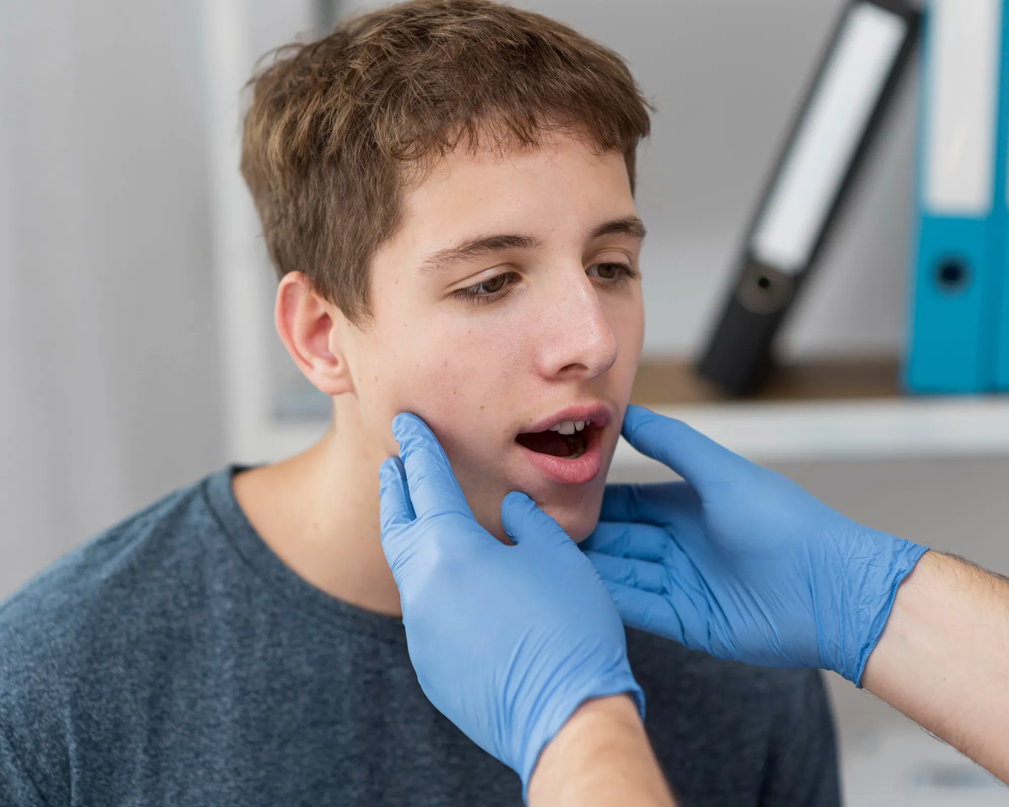
Diagnosis
The diagnosis of ameloblastoma typically begins with a thorough clinical evaluation, including a review of the patient’s medical history and a physical examination of the oral cavity and jaw. Imaging studies such as dental panoramic radiographs, X-rays, and CT scans are commonly used to visualize the extent and features of the tumor, including its size, place, and involvement of surrounding structures. In some cases, additional imaging modalities such as MRI or cone beam CT can be used to further evaluate the tumor and its relationship to adjacent tissues.
A definitive diagnosis of ameloblastoma is usually confirmed through a tissue biopsy, where a tiny sample of the tumor is collected and tested under a microscope by a pathologist. Histological analysis of the biopsy specimen can reveal characteristic features of ameloblastoma, such as the presence of epithelial islands, stellate reticulum-like cells, and peripheral palisading. Once diagnosed, further evaluation may be needed to determine the optimal treatment approach based on factors such as the dimension and place of the tumor, the presence of any associated complications or symptoms, and the patient’s overall health and treatment preferences.
Prevention
Preventing ameloblastoma is challenging due to its unclear etiology, but there are some general strategies that may help decrease the risk or find it early. Regular dental check-ups and routine dental X-rays can aid in the early detection of any abnormalities in the oral cavity, including potential signs of ameloblastoma. Additionally, maintaining good oral hygiene practices, such as brushing and flossing regularly and avoiding tobacco products, may help reduce the chance of certain oral health conditions that could potentially contribute to the development of ameloblastoma. Early detection and intervention are key in managing ameloblastoma effectively, so individuals with a family history of the condition or other risk factors may benefit from regular screenings and close monitoring by a dental or medical professional.
Summary
Ameloblastoma is a rare tumor of the jaw, typically diagnosed through clinical evaluation, imaging studies, and tissue biopsy. Risk factors include age, gender, genetic syndromes, and previous dental procedures. Treatment involves surgical resection, often supplemented with reconstruction and, in some cases, radiation therapy.
Regular dental check-ups and good oral hygiene practices may help with early detection and prevention. Prevention strategies focus on early detection through routine screenings and monitoring for individuals at risk. Early intervention is crucial for effective management and improved outcomes.