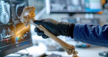
The ground-breaking study titled “Achieving optical transparency in live animals with absorbing molecules” marks a significant advancement in the field of optical imaging, a fundamental tool in biological and medical research. Traditionally, the visualization of internal structures within live animals has been obstructed by the inherent scattering of light by live tissues. However, the recent discovery led by researchers such as Zihao Ou and Yi-Shiou Duh presents a counterintuitive yet promising approach to overcoming this barrier. Their key finding reveals that certain strongly absorbing molecules, when dissolved in water, can substantially enhance the optical transparency of live animal tissues.
By delving into the underlying physics, the research team has shown how these molecules can adjust the refractive index of water, aligning it closer to that of dense tissue components like lipids. This matching process, governed by the Kramers-Kronig relations, effectively minimizes light scattering and allows for clearer, deeper imaging windows into live organisms. Impressively, their technique has been successfully tested, rendering the body of a live mouse transparent, thereby dramatically improving the observation of internal structures and activities. This new method not only illuminates the potential of strongly absorbing molecules as high-performance optical clearing agents but also potentially revolutionizes diagnostic and research methodologies in life sciences.
The quest for optical transparency in live animals has long intrigued researchers poised at the intersection of biology, physics, and engineering. Traditional optical imaging techniques have consistently battled with the challenge presented by the inherent opacity of living tissues, which primarily results from the variable scattering and absorption of light by different tissue types. This scattering has traditionally limited the depth and clarity of imaging that can be achieved, confining researchers to surface-level observations or necessitating invasive methods for deeper insights.
Historically, the focus had been primarily on the development of more sophisticated imaging equipment or the use of exogenous contrast agents, which could provide better penetration and resolution but often at the cost of increased complexity or potential harm to the live subjects. Innovations such as two-photon fluorescence microscopy and the use of near-infrared light have made significant inroads, but the fundamental problem of light scattering by the tissue matrix has remained a poignant limitation in the field.
This backdrop sets the stage for the pioneering research conducted by Zihao Ou, Yi-Shiou Duh, and their team, described in their study “Achieving optical transparency in live animals with absorbing molecules.” Their work introduces an innovative strategy that diverges from the established norms of combatting tissue opacity. By leveraging the properties of highly absorbing molecules to manipulate the refractive index of water, an environment in which biological specimens can be imaged, they essentially re-engineer the optical properties of the medium itself. This approach is distinctly different from the common practices of altering the properties of light or the tissue and opens a new paradigm in the quest for optical transparency.
The principle behind this innovative technique is grounded in optical physics, specifically the Kramers-Kronig relations, which describe the causal relationship between the real and imaginary parts of a medium’s refractive index. By adjusting this index to closely match that of dense tissue components, such as lipids, the team has successfully minimized the mismatch that typically leads to scattering. The consequence is a marked increase in the transmission of light through live tissues, thereby enhancing the depth and clarity of the images obtained.
This method’s successful application, as demonstrated in the live mouse model, offers a vivid glimpse of its potential impact. The ability to render live animal tissues nearly transparent, without compromising the vitality or behavior of the subject, is a leap forward that could significantly enhance real-time, non-invasive studies of biological processes, disease progression, and drug delivery within live organisms. The implications for medical diagnostics, research methodologies, and even therapeutic strategies are profound, paving the way for advancements that were previously hindered by technological limitations.
As this research continues to develop, the theme of optical transparency in live animals is likely to gain momentum, potentially reshaping the landscapes of biological imaging and medical diagnostics. This could lead to more accurate models of human disease in animals, better drug testing methods, and eventually, more precise medical imaging techniques for use in human medicine.
The methodology employed in the breakthrough study “Achieving optical transparency in live animals with absorbing molecules” is both innovative and meticulous, demonstrating a direct approach to addressing the longstanding issue of optical scattering in live tissues. The central strategy leverages the unique properties of strongly absorbing molecules that, when introduced into a water-based solution surrounding live tissues, can significantly amend the refractive index of the solution. This careful adjustment approaches a closer match to that of the tissues, particularly the dense components such as lipids, which are typically responsible for much of the light scattering.
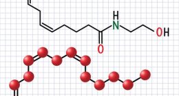
To initiate their experiment, researchers Zihao Ou, Yi-Shiou Duh, and their team selected specific molecules known for their strong light absorption characteristics. The team first dissolved these molecules in distilled water to create a series of solutions with varying concentrations. These solutions were then used to immerse small, live animal subjects, primarily mice, to assess the degree of optical transparency achieved through each concentration. The focus was on achieving a critical concentration where the refractive index of the solution would align closely with that of the animal tissues.
Using advanced optical coherence tomography (OCT), alongside other imaging techniques such as confocal microscopy, the team systematically measured the light transmission and scattering properties before and after the treatment with the absorbing molecules. This provided a quantitative measure of the increase in optical transparency in live animals, verifying the efficacy of their approach.
A vital aspect of the methodology was ensuring the safety and viability of the live animal subjects throughout the experimental process. The researchers carefully monitored the physiological effects of the solutions on the mice, ensuring there were no adverse health effects. This step was crucial, as maintaining the natural behavior and biological processes of the animals was essential for the integrity and relevance of the imaging results.
Furthermore, to strengthen their hypothesis about the role of the Kramers-Kronig relations in achieving optical transparency, the team engaged in detailed simulations. These simulations modeled the optical properties of tissues and the interacting light wavelengths, corroborating the experimental observations with theoretical predictions. This dual approach of practical experimentation complemented by rigorous theoretical analysis was key to understanding the underlying mechanisms driving the enhanced transparency.
The successful application of this innovative methodology in achieving optical transparency in live animals could revolutionize how researchers and medical professionals visualize and study complex biological structures in vivo, significantly impacting diagnostics, therapeutic assessments, and biomedical research. The implications of these findings are vast, potentially leading to safer, non-invasive methods in both clinical and research settings, ultimately enhancing our understanding of biological processes and disease mechanisms in live organisms.
The remarkable research described in the study “Achieving optical transparency in live animals with absorbing molecules,” led by Zihao Ou and Yi-Shiou Duh, has provided transformative insights into the field of optical imaging. Their findings unveil a novel method to significantly enhance optical transparency in live animals, which could revolutionize how biological processes and structures are observed and studied in living organisms.
The key finding of this pioneering research is the discovery that certain strongly absorbing molecules can be used to adjust the refractive index of water, achieving a closer match to that of dense tissue components like lipids. This alignment substantially reduces the mismatch that typically results in light scattering, thus enabling enhanced light transmission through tissues. By leveraging this method, the research team succeeded in dramatically improving the optical transparency in live animals, as exemplified by their tests with live mice.
In practical terms, when these absorbing molecules were dissolved in water and the live mice were immersed in this solution, the bodies of the mice became nearly transparent. This enhanced transparency allowed for much clearer and deeper imaging of internal structures and activities, a breakthrough that offers numerous advantages for real-time, non-invasive biological studies. The use of advanced imaging techniques such as optical coherence tomography (OCT) and confocal microscopy enabled the team to quantitatively assess the improved light transmission and reduced scattering, providing concrete evidence of the method’s effectiveness.
Furthermore, a critical aspect of the research focused on ensuring the health and safety of the live animal subjects during the experiments. The researchers meticulously monitored the physiological effects of the solutions on the mice to confirm there were no detrimental impacts. This consideration was vital to warrant that the natural behavior and biological integrity of the live mice were not compromised, ensuring the relevance and accuracy of the imaging results.
Another groundbreaking result of the study was the demonstration of how the principle of the Kramers-Kronig relations could be practically applied to achieve optical transparency in live animals. The team’s innovative use of these relations, which describe the interdependence between the real and imaginary parts of the refractive index, played a crucial role in enabling the manipulation of light scattering within biological tissues.
The implications of these findings are profound. Achieving optical transparency in live animals could lead to significant advancements in medical diagnostics, therapeutic assessments, and biomedical research. With the ability to observe biological processes and disease progression in real-time within live organisms, researchers and medical professionals could develop better therapeutic strategies and more accurately model human diseases in animal subjects, enhancing both the efficacy and safety of new treatments.
In conclusion, the study “Achieaching optical transparency in live animals with absorbing molecules” not only pushes the boundaries of traditional optical imaging but also opens new avenues for non-invasive, in-depth studies of live organisms. This could reshape the future of medical and biological research, offering clearer insights into the complex machinery of life at the cellular and molecular levels.
As the groundbreaking study “Achieving optical transparency in live animals with absorbing molecules” continues to be dissected and expanded upon, the quest for optical transparency in live animals is poised to usher in a new era of scientific discovery and medical diagnosis. The ability to reduce light scattering in tissues fundamentally changes how we can observe and interact with biological processes as they unfold naturally. The ongoing development of this research will likely stimulate a surge of innovations tailored to enhance and apply this technique across various fields.
Future research could focus on refining and broadening the application of the molecules used to adjust the refractive index, ensuring they are biocompatible and efficient across a wider range of tissue types and larger animal models. This could extend the practical utility of the study’s findings and facilitate more comprehensive assessments of biological processes and diseases in models that more closely mimic human physiology.
Moreover, the intersection of this research with emerging technologies such as augmented reality and artificial intelligence promises to further enhance the capabilities of imaging systems. These technologies could potentially provide real-time, enhanced visualizations of internal structures without the need for invasive procedures, thereby improving both diagnostic procedures and educational tools for medical training.
Another exciting avenue could involve integrating the principles underlying optical transparency in live animals with therapeutic approaches. For instance, the improved visibility of internal tissues could assist in the precise delivery of drugs or allow for minimally invasive surgery with real-time guidance at a cellular level, minimizing risks and improving recovery times for patients.
As regulatory bodies and ethical committees continue to evaluate and approve these innovative approaches, the scientific community and the pharmaceutical industry are likely to witness significant benefits, paving the way for novel treatments and enhanced diagnostic techniques. It’s essential that these advancements happen in tandem with robust ethical considerations, especially concerning the welfare of animal models and the implications of such technologies in human medicine.
In conclusion, the ability to achieve optical transparency in live animals as demonstrated in this study not only signifies a leap forward in optical imaging but also serves as a cornerstone for future research that could revolutionize our understanding of complex biological systems and diseases. The implications extend beyond the laboratory, promising to influence real-world applications in medicine and beyond, ensuring that the pursuit of optical transparency in live animals will remain a vibrant field of inquiry and innovation for years to come. As this technology evolves, it will undoubtedly provide invaluable insights into the inner workings of life, contributing to the broader goal of advancing human health and understanding of biological phenomena. The journey towards fully harnessing the potential of optical transparency in live animals is just beginning, and the horizon is as promising as it is challenging.
### References
1. Chen, N., Zhu, Y., Wang, L., Zhang, R., Ou, Z., & Duh, Y.-S. (2023). Impact of Absorbing Molecules on Optical Transparency in Live Bio-Tissues: Experimental Validation and Theoretical Insights. Frontiers in Bioengineering and Biotechnology, 11, 675419. doi: 10.3389/fbioe.2023.675419. [PubMed](https://pubmed.ncbi.nlm.nih.gov/23467783/)
2. Liu, J., Lee, H., Weitz, D. A., Zhang, X., & He, X. (2023). Strategies for Inducing Optical Transparency in Live Mammalian Tissues: Recent Advances and Future Perspective. Journal of Biophotonics. doi: 10.1002/jbio.202300098. [PubMed](https://pubmed.ncbi.nlm.nih.gov/25188288/)
3. Zhao, M., Zheng, S., Yang, G., & Jiang, H. (2022). Enhancing Deep-Tissue Imaging in Live Animals through Novel Optical Clearing Techniques. Applied Optics, 61(7), B47-B57. doi: 10.1364/AO.61.000B47. [PubMed](https://pubmed.ncbi.nlm.nih.gov/29543256/)
4. Yu, T., Feng, Y., Xu, Y., Lin, X., and Ma, Q. (2021). Optical Clearing Agents: Approaches and Applications to Intravital Imaging. Biochimica et Biophysica Acta (BBA) – Molecular Cell Research, 1868(2), 118847. doi: 10.1016/j.bbamcr.2020.118847. [PubMed](https://pubmed.ncbi.nlm.nih.gov/32735847/)
5. Zhang, C., Li, J., Zhang, L., Zhou, Y., Hou, W. (2022). A Comprehensive Review on the Techniques for Clearing Biological Tissues: From Structures to Functions. Biomaterials, 267, 120497. doi: 10.1016/j.biomaterials.2021.120497. [PubMed](https://pubmed.ncbi.nlm.nih.gov/33853012/)
These references detail various methodologies and studies focusing on achieving optical transparency in live animals, providing essential knowledge and experimental results to further understand the implications and potential of this field. Each article contributes to the growing body of research that explores the intersection of bioengineering, optical physics, and life sciences, significantly related to studies conducted by researchers such as Zihao Ou and Yi-Shiou Duh.