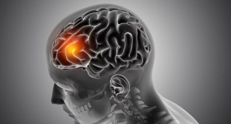
Cysts can develop in any part of the body, as well as the brain. Many brain cysts are harmless and usually don’t need surgery. If surgery is needed, the doctor may shrink or eliminate the cyst.
A cyst is a pouch filled with a substance like fluid or air. It may look like a tumor on the outside, but they are caused differently. A tumor is a lump of tissue, while a cyst is a pouch filled with fluid.
Causes
The causes of brain cysts can include:
- Congenital: Some cysts are present from birth due to developmental abnormalities.
- Infection: Certain infections can lead to the formation of brain cysts.
- Inflammatory conditions: Conditions that cause inflammation in the brain may result in cyst formation.
- Parasitic infestations: In rare cases, parasites can cause cysts to form in the brain.
- Tumors: Some tumors can degenerate and form cysts within the brain tissue.
Types, Symptoms, and Treatment
Arachnoid Cysts
Arachnoid cysts grow on the arachnoid membrane, which covers the spinal cord and brain alongside the other two membranes. These cysts are generally non-cancerous and often don’t cause signs. However, when symptoms do occur, they can include seizures, headaches, nausea, vomiting, and dizziness. In cases of spinal arachnoid cysts, symptoms may manifest as a lack of sensation in the hands or continuous weakness in the legs.
Since most arachnoid cysts are asymptomatic, healthcare providers typically detect them incidentally during CT or MRI scans conducted for other reasons. Treatment is usually unnecessary except if the cyst shows symptoms. If intervention is necessary, surgery may be performed to shrink the fluid from the cyst, which can then be absorbed by nearby tissue.
Pineal Gland Cysts
These are fluid-contained spaces found in the pineal gland, which controls sleep cycles and is located near the brain’s center. Many pineal cysts are non-cancerous and often produce minimal or no signs. However, larger cysts, exceeding 2 cm in size, may lead to headaches, vertigo, or visual troubles, according to older medical sources.
Surgical removal of pineal cysts is uncommon unless they significantly impact brain function or cause symptoms that affect daily life. When necessary, the procedure involves creating a small opening in the cyst to drain fluid, which then flows into the brain’s substance gaps.
Colloid Cysts
These cysts grow within the brain’s ventricles, which are fluid-filled cavities containing cerebrospinal fluid. Although benign, these cysts can obstruct cerebrospinal fluid flow, resulting in a condition known as hydrocephalus. Signs of hydrocephalus include memory problems, headaches, trouble concentrating, double vision, and in severe cases, consciousness or coma.
Healthcare providers typically treat colloid cysts using minimally intrusive procedures such as endoscopy, which allows them to eliminate the cyst and alleviate hydrocephalus by restoring normal fluid flow within the brain.
Dermoid Cysts
These cysts originate from a congenital malfunction in skin cell development, where layers of skin fail to fuse properly during fetal growth. These cysts can contain hair follicles, sweat glands, oil glands, and occasionally, teeth, bone, or cells of nerves. While dermoid cysts are generally non-cancerous and often asymptomatic, they are usually surgically removed shortly after birth to prevent potential complications.
Epidermoid Cysts
Epidermoid cysts differ from dermoid cysts in that they carry simple skin cells, involving dead skin cells and keratin. Found in the spine, these cysts develop when the body eliminates inside skin cells, resulting in slow cyst growth. While typically not dangerous on their own, epidermoid cysts can compress the nerves or spinal cord, leading to symptoms such as weakness, decreased coordination, tingling, incontinence, and mobility issues.
Treatment for epidermoid cysts typically involves microsurgery techniques to safely remove the cyst and alleviate pressure on the nerves or spinal cord, restoring normal function.
Complications
Brain cysts, depending on their kind and position, can lead to different complications:
- Increased Intracranial Pressure: Sometimes brain cysts block the flow of cerebrospinal fluid, leading to increased pressure in the skull internally. This condition, known as hydrocephalus, can lead to headaches, nausea, throwing up, and, in serious cases, neurological signs like altered consciousness.
- Compression of Nearby Structures: Large cysts can compress nerves, adjacent brain tissue, or blood vessels. This compression can result in neurological deficits like numbness, weakness, impaired coordination, or sensory disturbances.
- Symptomatic Epilepsy: Certain kinds of brain cysts, particularly those situated in areas responsible for controlling neurological activity, can active seizures.
- Rupture or Hemorrhage: In uncommon cases, cysts may bleed or rupture, cause sudden onset of serious headaches, neurological deficits, or even dangerous conditions like intracranial hemorrhage.
- Cancerous Transformation: Although most brain cysts are non-cancerous, in some cases, there can be a chance of cancerous transformation, where the cystic structure may turn cancerous.
- Infection: Cysts can sometimes become infected, leading to signs like worsening headaches, fever, and neurological deficits.
- Functional disability: Depending on their location, cysts can interfere with normal brain function, leading to cognitive impairment, memory issues, or changes in mood and behavior.
Early identification and suitable management by healthcare providers are crucial in minimizing these potential complications connected with brain cysts.
Prevention
Preventing brain cysts is largely based on the underlying causes, many of which are congenital or connected to developmental malformation. Since many brain cysts are not avoidable through lifestyle modifications or particular actions, early diagnosis and monitoring are key. Regular medical check-ups, specifically for people with a family history of neurological conditions or known genetic predispositions, can aid in early detection.
Timely medical treatment for conditions that may predispose to cyst formation, like infections or inflammatory disorders affecting the brain, can also help handle risks. Overall, continuing a healthy lifestyle with a nutritious diet, regular physical activities, and avoiding risk factors like head injuries or untreated infections, can support overall brain health, potentially reducing the possibility of issues related to cyst formation.
Summary
Brain cysts vary in type and location, including arachnoid, pineal gland, colloid, dermoid, and epidermoid cysts, each with distinct characteristics and potential symptoms such as headaches and neurological deficits. While most are benign and may not require treatment unless symptomatic, complications like hydrocephalus or neurological impairment can arise if untreated.
Surgical intervention, often through minimally invasive techniques, may be necessary for symptomatic cysts to alleviate pressure or restore normal brain function. Prevention strategies focus on early detection through regular medical check-ups, especially for those with genetic predispositions or neurological conditions. Maintaining a healthy lifestyle and promptly treating conditions that may lead to cyst formation is crucial in managing risks associated with brain cysts.


